

Jun-Won Lee, Seung-Hwan Lee, et al.
[Myocardial Infarction Phase 3] Safety and Effects on Cardiac Function of Coronary Artery Injection of Autologous Bone Marrow-Derived Mesenchymal Stem Cells in Patients with Acute Myocardial Infarction (SEED-MSC Study)
Medium-sized stem cells cultured from the patients’ own bone marrow were administered into the coronary arteries of 58 patients who had undergone reperfusion therapy for acute myocardial infarction. Six months later, the left ventricular ejection fraction—a measure of the heart’s pumping function—increased by 5.9 percentage points in the treatment group and by 1.6 percentage points in the control group, and there were no serious complications related to the procedure. However, there were no changes in heart size or wall motion, and given the small number of participants, it is too early to generalize these results.
Study Results
Approximately one month after patients with acute myocardial infarction underwent reperfusion therapy to reopen blocked blood vessels, mesenchymal stem cells cultured from their own bone marrow were administered into their coronary arteries. The primary endpoint was the change in left ventricular ejection fraction (LVEF; the percentage of blood ejected by the heart with each contraction) at the 6-month mark.
SPECT scans showed that the ejection fraction in the treatment group increased by an average of 5.9 percentage points (±8.5), while that in the control group increased by 1.6 percentage points (±7.0) (P = 0.037). Echocardiography also showed a difference, with the treatment group showing a 1.9 percentage point increase and the control group a 0.5 percentage point decrease (P < 0.001). However, there was no difference between the two groups in left ventricular volume or wall motion index. When analyzing only patients who received reperfusion therapy within 6 hours of symptom onset, the difference was even greater (8.3 percentage points in the treatment group vs. 1.3 percentage points in the control group based on SPECT, P = 0.007).
In terms of safety, both the bone marrow collection and cell infusion procedures proceeded without major issues. Two patients experienced elevated cardiac enzymes immediately after administration but recovered without symptoms. During the 6-month follow-up period, there were no cases of death, reinfarction, target vessel revascularization, stent thrombosis, fatal arrhythmia, or stroke in either group.
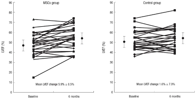
Figure. Changes in left ventricular ejection fraction in the treatment and control groups (baseline → 6 months). Original Figure 2, J Korean Med Sci.
Study Design
This was a randomized, open-label, multicenter clinical trial conducted at three tertiary general hospitals in Korea from March 2007 to September 2010 (ClinicalTrials.gov NCT01392105). Eighty patients aged 18–70 who had successfully undergone reperfusion therapy for ST-segment elevation myocardial infarction were screened; 69 were assigned to the treatment group (33 patients) and the control group (36 patients). The final analysis included 58 patients(30 in the treatment group and 28 in the control group) were included in the final analysis. The control group received only standard medical therapy and did not undergo a sham procedure.
The primary endpoint was the change in left ventricular ejection fraction (LVEF) measured by SPECT at 6 months, and secondary endpoints included left ventricular volume, wall motion index, and major cardiac events. Follow-up evaluations were conducted at 1 month, 2 months, and 6 months, and included coronary angiography, SPECT, echocardiography, and 24-hour Holter monitoring.
Cells Used and Administration Method
Autologous bone marrow-derived mesenchymal stem cells obtained from the patients’ own bone marrow were used. On average, 20–25 mL of bone marrow was collected from the posterior iliac crest 3.8 days after hospitalization and cultured to 4–5 passages in a GMP facility; the average time from collection to administration was 25 days. The release criteria were a viability rate of 80% or higher, negative for microbial contamination, at least 90% of cells positive for CD73 and CD105, and less than 3% of cells positive for CD14, CD34, and CD45.
The dosage was 1 million cells per kilogram of body weight, averaging 7.2 × 10⁷ cells per patient. A balloon catheter was inserted into the occluded coronary artery, and the balloon was inflated at low pressure from inside the stent to block blood flow for 2–3 minutes, during which the cells were infused. Blood flow was restored for 3 minutes at a time, and this process was repeated three times.
Discussion
This study demonstrates that the administration of autologous bone marrow-derived mesenchymal stem cells into the coronary arteries approximately one month after an acute myocardial infarction is feasible without major complications and resulted in a modest improvement in left ventricular ejection fraction at the 6-month mark. The magnitude of improvement is comparable to the 2.99% reported in a previous meta-analysis of bone marrow cell therapies. The research team noted that it is still difficult to determine what this level of improvement in contractile function means for patients’ daily lives. It is also important to consider that left ventricular volume and wall motion index remained unchanged.
Regarding the timing of administration, the researchers explained that the period after the acute inflammatory response has subsided but before scar tissue has solidified may be favorable for cell therapy.
There are also clear limitations. The number of participating patients was small, and the study used an open-label design without a placebo procedure in the control group. Due to issues such as poor image quality, 22 out of 80 participants (27.5%) were excluded from the analysis. Cardiac function was measured using SPECT instead of cardiac MRI, and indicators such as walking distance or quality of life were not evaluated. The fact that the participating patients were a low-risk group with relatively well-preserved cardiac function, and that the observation period was limited to 6 months, must also be taken into account in interpreting the results. This study was conducted with research funding from Pamcell.


